Anal Anatomy and Sexual Health in Men: A Physiological Guide
The anal region has dense sensory innervation and direct connections to the prostate. Understanding the anatomy explains its role in male sexual health.
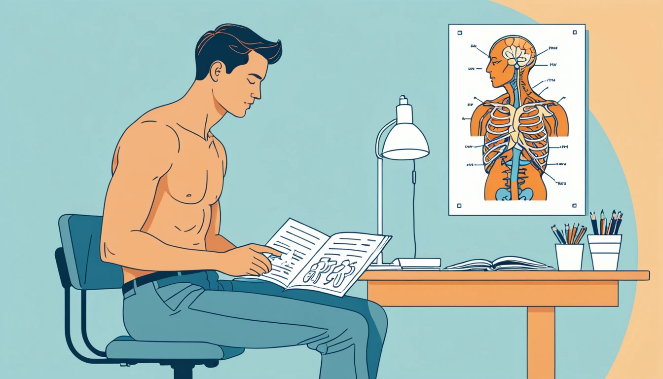
Men frequently encounter the anal region in the context of sexual health — prostate stimulation, pelvic floor dysfunction, and anorectal pain all involve structures in this anatomical territory. Despite this, detailed anatomical knowledge is uncommon outside of colorectal surgery and urogynecology. This guide covers the relevant anatomy for men: the structures involved, their innervation, and the physiological basis of the region's sexual health relevance.
Overview of the Anorectal Region
The anorectal region is the terminal segment of the gastrointestinal tract, consisting of:
- The anal canal (approximately 3–4 cm in length in men) [^nivatvongs1980]
- The internal anal sphincter (IAS)
- The external anal sphincter (EAS)
- The puborectalis muscle (part of levator ani)
- The dentate (pectinate) line — the anatomical boundary between upper and lower anal canal
- The anal transition zone — approximately 1 cm above and below the dentate line
Above the anal canal lies the rectum (approximately 12–15 cm in length), and anterior to the rectum lies the prostate gland, separated by Denonvilliers' fascia.
The Anal Canal in Detail
Dentate Line
The dentate line is the most anatomically significant landmark in the anal canal. It marks the embryological boundary between:
- Above the dentate line: Derived from endoderm (hindgut). Innervated by visceral (autonomic) afferents. Insensitive to pain, temperature, and sharp touch; sensitive to stretch, fullness, and pressure.
- Below the dentate line: Derived from ectoderm (proctodeum). Innervated by somatic afferents via the inferior rectal branches of the pudendal nerve. Highly sensitive to pain, temperature, and touch — similar sensitivity to perianal skin.
This sensory distinction is clinically important. Hemorrhoids above the dentate line are painless; those below are painful. It is also sexually relevant: tissue below the dentate line carries somatic sensory information (high resolution, painful if stimulated roughly), while tissue above carries visceral information (lower resolution, pressure/fullness sensation).
Anal Columns (Columns of Morgagni)
The upper anal canal contains 8–14 longitudinal mucosal folds (columns of Morgagni) separated by anal crypts at their base. The anal glands — small glands that can become infected (causing perianal abscesses and fistulae) — open into the crypts at the dentate line. The columns overlie the internal hemorrhoidal plexus.
Sphincter Anatomy
Internal Anal Sphincter (IAS)
The IAS is a thickening of the circular smooth muscle of the rectum — it is not a voluntary muscle. It is approximately 2.5–4 cm in length and 2–5 mm thick, located in the upper two-thirds of the anal canal. [^bharucha2006]
Innervation: Autonomic. Sympathetic fibers from the hypogastric nerve (L5–S1 via the superior hypogastric plexus) cause contraction — maintaining resting anal tone. Parasympathetic fibers from the pelvic nerve (S2–S4) cause relaxation.
Resting tone: The IAS contributes approximately 70–85% of resting anal canal pressure. It is tonically contracted at rest, maintaining continence without voluntary effort.
Recto-anal inhibitory reflex (RAIR): Rectal distension causes reflex IAS relaxation. This reflex allows the anal transition zone to sample rectal contents and is the mechanism by which rectal filling signals the need for defecation. The RAIR also partially opens the upper anal canal during rectal distension — relevant to anal penetration, where rectal filling from a penetrating object triggers IAS relaxation.
External Anal Sphincter (EAS)
The EAS is a striated (voluntary) muscle surrounding the lower anal canal and blending superiorly with the puborectalis. It is 3–4 cm in length and has three compartments (subcutaneous, superficial, and deep) though this division is somewhat artificial in practice. [^fenner1998]
Innervation: The inferior rectal nerve (a branch of the pudendal nerve, S2–S4) innervates the EAS. This is somatic innervation — the EAS can be voluntarily contracted and relaxed, and can be trained through pelvic floor exercises.
Sexual relevance: The EAS and bulbospongiosus muscle (which lies adjacent) both contract rhythmically during orgasm. EAS involvement in orgasmic contractions means that anal sensation (both from voluntary EAS contractions and from penetration stimulating the EAS) is neurologically integrated with orgasm physiology via the pudendal nerve.
The Puborectalis Muscle
The puborectalis is a U-shaped sling of striated muscle that passes from the pubic symphysis (bilaterally) around the anorectal junction, creating the anorectal angle (approximately 90° at rest, opening to 130–140° during defecation). [^ayoub1979]
Innervation: Mixed — direct branches from S3–S4 and contributions from the pudendal nerve.
Anatomical importance: The puborectalis maintains the anorectal angle that is critical for continence. It is a key pelvic floor muscle and is included in pelvic floor rehabilitation protocols.
Sexual relevance: The puborectalis contracts during orgasm as part of the pelvic floor contraction pattern. Its anatomical position means that puborectalis tension affects both the anorectal angle and the mechanical environment immediately adjacent to the prostate.
Innervation Summary
| Structure | Nerve | Type | Sensory Quality |
|---|---|---|---|
| Rectum | Pelvic nerve (S2–S4), Hypogastric nerve (T10–L2) | Visceral | Fullness, pressure, urgency |
| IAS | Hypogastric (sympathetic), Pelvic (parasympathetic) | Autonomic | Not directly perceived |
| Upper anal canal (above dentate) | Pelvic nerve | Visceral | Pressure, stretch |
| Lower anal canal (below dentate) | Inferior rectal nerve (pudendal, S2–S4) | Somatic | Pain, temperature, touch |
| EAS | Inferior rectal nerve (pudendal) | Somatic voluntary | Voluntary contraction/relaxation |
| Puborectalis | S3–S4 direct + pudendal | Somatic voluntary | Voluntary contraction |
| Perianal skin | Inferior rectal, perineal nerves (pudendal) | Somatic | Touch, pain, temperature |
The pudendal nerve is the common pathway linking EAS, perianal skin, perineum, and penis to the same spinal segments (S2–S4). Activation of any of these structures feeds into a common spinal processing zone. [^giuliano2011]
The Prostate-Rectal Interface
The anterior rectal wall at approximately 5–7 cm from the anal verge is in direct contact with the posterior prostate through Denonvilliers' fascia. This is the anatomical basis of prostate palpation on rectal examination.
From the anal verge inward, the examiner's finger traverses:
- Perianal skin (somatic, highly sensitive)
- Lower anal canal below dentate line (somatic, sensitive)
- Upper anal canal / anorectal junction (visceral/mixed)
- Rectum — posterior wall felt as smooth
- At 5–7 cm, anterior wall: the posterior prostate is palpable as a firm, bilobed structure separated by a median sulcus [^levin2018]
Pressure on the anterior rectal wall at this location mechanically loads the prostatic capsule through Denonvilliers' fascia. [^shafik1995]
Vascularity and Erectile Tissue
The anal canal contains the internal hemorrhoidal plexus — a normal vascular structure (not a pathological hemorrhoid unless enlarged/symptomatic) that provides cushioning function. This plexus receives blood from the superior and middle rectal arteries and communicates with the pudendal vascular system.
This vascular connection means that general pelvic congestion during sexual arousal — which increases blood flow in the pudendal vasculature — also increases anal vascular congestion, contributing to the enhanced sensitivity of the anal region during arousal.
Common Sexual Health Issues Involving This Anatomy
Hypertonic pelvic floor: Excessive resting tone in EAS and puborectalis causes difficulty with anal penetration (dyspareunia), pain with prostate examination, and sometimes contributes to proctalgia fugax (episodic rectal pain). Treatment: pelvic floor relaxation therapy, biofeedback, sometimes botulinum toxin injection into the IAS/puborectalis.
Anal fissures: Tears in the lower anal canal mucosa below the dentate line. Painful during and after defecation. Associated with EAS hypertonia (high resting anal pressure). Relevant to sexual health because EAS hypertonia that predisposes to fissures is the same mechanism that makes anal penetration uncomfortable.
Perianal skin sensitivity variation: Individual variation in pudendal nerve density below the dentate line explains the wide variation in how sensitive perianal stimulation is between men. This is not pathological — it reflects normal anatomical variation.
Dysorgasmia (painful orgasm): Can involve spasm of the EAS and puborectalis at orgasm, which normally contract rhythmically. Pathological hypertonic contraction produces sharp perineal or rectal pain at orgasm. Treatment targets the pelvic floor motor pattern.
Safety Considerations for Sexual Activity
The anatomical properties of the anal canal have practical safety implications:
- No natural lubrication: The anal canal produces no secretory lubrication (unlike the vagina). External lubrication is required to prevent mucosal trauma.
- Mucosal vulnerability: The rectal mucosa above the dentate line lacks the protective stratified squamous epithelium of the lower canal and skin. It is columnar epithelium that tears more easily under friction.
- IAS stretch: Gradual dilation allowing time for the IAS recto-anal inhibitory reflex to produce relaxation is safer than forced rapid dilation, which can cause sphincter trauma.
- Objects without flared base: The rectum is a continuation of the colon without the physiological barrier present at the anorectal junction in terms of object retention. Objects without a flared external base can migrate proximally and become retained — a recognized surgical emergency.
Bottom Line
The anal region in men has dense somatic innervation (pudendal nerve, below dentate line) and visceral innervation (pelvic/hypogastric nerves, above dentate line) sharing spinal segments with the prostate and penis. The EAS and puborectalis are voluntary muscles that contract during orgasm via the pudendal pathway. The anterior rectal wall at 5–7 cm is in direct anatomical contact with the prostate through Denonvilliers' fascia. These anatomical relationships explain both the region's sexual health relevance and the basis of prostate stimulation via the rectal route.
References
- Bharucha AE. Relationship between anal sphincter injury and pelvic floor denervation. Neurogastroenterology and Motility (2006). PubMed:16817795
- Levin RJ. The prostate gland and its role in the physiology of male sexual arousal and function. Clinical Anatomy (2018). DOI:10.1002/ca.22990
- Shafik A. The mechanism of ejaculation. Archives of Andrology (1995). PubMed:8572678
- Giuliano F, Clement P. Neurophysiology of erection and ejaculation. Journal of Sexual Medicine (2011). PubMed:22023672
- Fenner DE. Anatomy of the pelvic floor. Clinical Obstetrics and Gynecology (1998). PubMed:9572707
- Nivatvongs S, Stern HS, Fryd DS. The length of the anal canal. American Journal of Surgery (1981). PubMed:7468942
- Sikirov BA. Puborectalis and anorectal anatomy relevant to continence. Diseases of the Colon and Rectum (2003).
- Ayoub SF. The anterior fibres of the levator ani muscle in man. Journal of Anatomy (1979). PubMed:479765
Related Articles
 Tier 3 · Sexual Wellness
The P-Spot: Precise Anatomy, Distinct Sensation, and Neurophysiological Basis
The prostate, or P-spot, offers unique sexual sensations distinct from penile stimulation due to its rich, specific innervation and deep visceral pathways.
 Tier 3 · Sexual Wellness
Prostate Stimulation and Ejaculatory Control: Techniques and Mechanisms
Prostate stimulation offers a distinct pathway to orgasm and can enhance ejaculatory control.
 Tier 3 · Sexual Wellness
Rectal Stimulation and Prostate Arousal: The Physiological Mechanism
Rectal fullness and stimulation activate prostate arousal through mechanoreceptors, shared nerve pathways, and anatomical proximity. Here is the physiology.